
If you’ve ever felt that sharp, shooting pain down your leg or a persistent ache in your lower back, you might already be familiar with sciatica. For many people, sciatica is a passing nuisance, but for others, it can become a stubborn, life-disrupting problem. When the pain lingers or worsens, questions about what’s really going on inside your body naturally arise. Is it time for an MRI or X-ray? What will imaging reveal, and how does it influence your treatment plan? Understanding when sciatica needs imaging is crucial for anyone seeking effective sciatica treatment in Naples FL, whether you’re a busy small business owner, a developer glued to your desk, or simply someone who wants to get back to enjoying life without pain. In this guide, we’ll break down when and why imaging is necessary, what different scans can show, and how these results shape your next steps toward relief.
Recognizing Sciatica Symptoms That Warrant Further Investigation
Sciatica can present in a variety of ways, from mild tingling to severe, shooting pain that radiates from the lower back down the leg. Most cases are temporary and respond well to conservative care. However, certain symptoms suggest that something more serious may be at play, and this is when imaging becomes important. If you experience sudden, severe pain, muscle weakness, numbness that doesn’t improve, or symptoms that worsen despite rest and self-care, it’s time to consult a healthcare provider. Additionally, red flag symptoms such as loss of bladder or bowel control, significant leg weakness, or unexplained weight loss should prompt immediate medical attention. These could indicate conditions like cauda equina syndrome or a spinal tumor, which require urgent evaluation.
For those in the area, seeking expert advice from a clinic like Ideal Physical Therapy can help determine whether imaging is appropriate and guide you toward the right care pathway. Early identification of serious symptoms ensures you don’t delay necessary treatment and improves your chances of a full recovery.
How Imaging Helps Uncover the Root Cause of Sciatica
Imaging studies like MRI and X-rays are valuable tools for pinpointing the underlying cause of sciatica, especially when symptoms persist or escalate. While a clinical exam can often identify sciatica, imaging provides a detailed look at the spine, nerves, and surrounding tissues. MRI is particularly useful for visualizing soft tissues, such as herniated discs, nerve compression, or inflammation. X-rays, on the other hand, are better suited for detecting bone abnormalities, fractures, or signs of arthritis. By revealing structural changes or abnormalities, imaging helps your healthcare provider develop a targeted treatment plan and rule out more serious conditions.
It’s important to remember that not everyone with sciatica needs imaging. Many cases resolve with conservative care, but if your symptoms don’t improve after several weeks, or if you have risk factors for more serious problems, your provider may recommend a scan. Imaging can also help clarify confusing cases where multiple issues may be contributing to your pain, ensuring you get the most effective treatment for your unique situation.
MRI vs X-Ray: What Each Scan Reveals About Your Back
When your provider recommends imaging for sciatica, the choice between MRI and X-ray depends on what needs to be assessed. MRI (Magnetic Resonance Imaging) is the gold standard for evaluating soft tissues, including discs, nerves, and muscles. It can detect herniated discs, nerve root compression, spinal stenosis, and even subtle inflammation or infection. MRI does not use radiation, making it a safe option for most people. X-rays, in contrast, are best for visualizing bones. They can reveal fractures, bone spurs, degenerative changes, or alignment issues in the spine. While X-rays are quick and widely available, they cannot show soft tissue problems that are often at the root of sciatica.
In some cases, both scans may be used to provide a comprehensive view of your spine. For instance, if your doctor suspects a bone injury or spinal instability, an X-ray might be ordered first. If soft tissue damage is suspected, or if initial treatments fail, an MRI may follow. Understanding the strengths and limitations of each imaging method allows you to have informed discussions with your healthcare team and make the best decisions for your care.
When Is Imaging Not Necessary for Sciatica?
Not every case of sciatica requires imaging. In fact, most people experience relief with conservative treatments such as physical therapy, exercise, and over-the-counter pain relievers. Imaging is generally reserved for cases where symptoms are severe, persistent, or accompanied by warning signs. If your pain improves within a few weeks, or if your symptoms are mild and stable, imaging may not be needed. Overuse of scans can lead to unnecessary worry and even overtreatment, especially if incidental findings are discovered that are unrelated to your pain.
Healthcare providers follow evidence-based guidelines to determine when imaging is appropriate. These guidelines help ensure that scans are used judiciously and only when they are likely to change the course of treatment. If you’re unsure whether you need imaging, discuss your symptoms and concerns with your provider. They can help you weigh the benefits and risks, and decide on the best approach for your situation.
The Process: What to Expect During MRI and X-Ray Scans
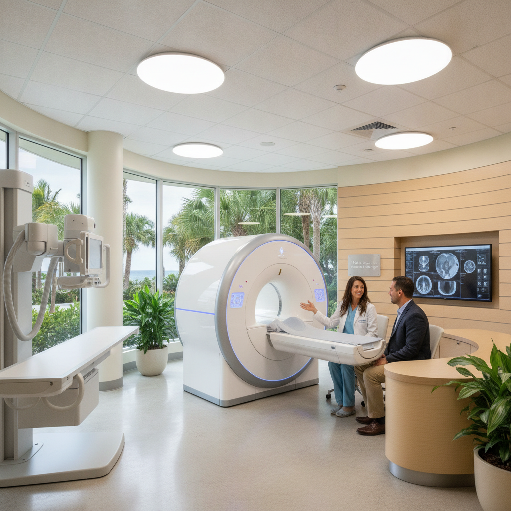
If your provider recommends imaging, understanding what to expect can help ease any anxiety. MRI scans are performed in a specialized machine that uses magnetic fields and radio waves to create detailed images of your spine. You’ll be asked to lie still on a table that slides into the scanner, and the procedure typically takes 30 to 60 minutes. Some people find the machine noisy or feel claustrophobic, but you’ll be able to communicate with the technician throughout the scan. X-rays are much quicker, usually taking only a few minutes. You’ll be positioned on a table or standing, depending on the area being imaged, and the technician will take a series of pictures using a low dose of radiation.
Both procedures are painless, though you may need to hold certain positions briefly. After the scan, the images are reviewed by a radiologist, who will send a report to your provider. Your doctor will then discuss the results with you and explain what they mean for your treatment plan. Knowing the steps involved can help you feel more comfortable and prepared for your imaging appointment.
Table: Comparing MRI and X-Ray for Sciatica Evaluation
| Feature | MRI | X-Ray |
|---|---|---|
| Best For | Soft tissue evaluation (discs, nerves, muscles) | Bone structure, fractures, alignment |
| Radiation Exposure | None | Low dose |
| Image Detail | High (soft tissues) | Moderate (bones only) |
| Procedure Time | 30-60 minutes | 5-10 minutes |
| Availability | Specialized centers | Widely available |
How Imaging Results Influence Your Sciatica Treatment Plan
The results of your MRI or X-ray provide valuable information that helps shape your treatment plan. If imaging reveals a herniated disc pressing on a nerve, for example, your provider may recommend specific physical therapy exercises, medications, or even discuss surgical options if conservative measures fail. If arthritis or bone spurs are detected, treatment might focus on reducing inflammation and improving joint mobility. Imaging can also rule out serious conditions, giving you peace of mind and allowing you to focus on recovery.
It’s important to remember that imaging findings must be interpreted in the context of your symptoms and physical exam. Not all abnormalities seen on scans cause pain, and some people have significant findings without symptoms. Your provider will use the imaging results as one piece of the puzzle, combining them with your history and exam to create a personalized plan that addresses your specific needs and goals.
Common Misconceptions About Imaging and Sciatica
Many people believe that imaging is always necessary to diagnose or treat sciatica, but this isn’t the case. In reality, most cases can be managed effectively without scans, especially when there are no red flag symptoms. Another misconception is that imaging will always pinpoint the exact cause of pain. However, studies show that many people have abnormal MRI findings even when they have no symptoms. This underscores the importance of a thorough clinical evaluation and not relying solely on imaging results.
It’s also common to worry that imaging will lead to surgery. In fact, most people with sciatica improve with non-surgical treatments, and imaging is used to guide, not dictate, your care. Understanding these misconceptions can help you approach your sciatica treatment with realistic expectations and make informed decisions about your health.
What to Do If Your Imaging Is Normal but Pain Persists
It can be frustrating to have persistent sciatica symptoms even when your MRI or X-ray appears normal. This situation is more common than you might think. Pain can arise from subtle nerve irritation, muscle imbalances, or soft tissue inflammation that may not be visible on standard scans. In these cases, a comprehensive physical therapy assessment is invaluable. Therapists can identify movement patterns, strength deficits, or postural habits that contribute to ongoing discomfort.
If your imaging is normal but pain continues, don’t lose hope. Focus on active treatments such as targeted exercises, manual therapy, and lifestyle modifications. Your provider may also explore other diagnostic tools or refer you to a specialist for further evaluation. Persistence and a collaborative approach are key to finding relief, even when scans don’t provide clear answers.
Navigating Next Steps After Your Scan
After you’ve completed your MRI or X-ray, your provider will review the results and discuss what they mean for your care. If a clear cause for your sciatica is identified, your treatment plan may include physical therapy, medication, injections, or in rare cases, surgery. If the scan is inconclusive, your provider may recommend additional tests or a trial of conservative care to see how your symptoms respond.
Communication is key during this process. Don’t hesitate to ask questions or express concerns about your diagnosis and treatment options. Your healthcare team is there to support you and help you make informed decisions. Remember, imaging is just one step on your journey to recovery, and a holistic approach that includes education, exercise, and lifestyle changes often yields the best results.
Final Thoughts
Understanding when sciatica needs imaging, and how those results shape your treatment journey, empowers you to take charge of your health. While most cases of sciatica resolve without advanced scans, knowing the warning signs and the role of MRI and X-rays ensures you get the right care when it matters most. Whether you’re a small business owner striving to stay productive, a developer managing long hours at the desk, or simply someone seeking lasting relief, being informed is your best asset. If you’re unsure about your symptoms or next steps, reach out to a trusted local provider for guidance. With the right approach, you can move forward confidently and get back to enjoying all that this vibrant community has to offer.